Timed Pregnant Mice: Practical Tips for Improving Success
Need embryos or pups at a specific developmental stage? Timed matings are one of the most effective tools for generating age-matched cohorts and planning experiments with greater confidence. Evaluating a large number of aged-matched embryos or neonates likely will require setting up timed matings. Timed mating in mice facilitates experimental planning by allowing for predictable availability of embryos or pups, generation of age-matched experimental cohorts, and early identification of successful matings. Timed mating in mice enables precise determination of embryonic age, with the day a vaginal plug is detected typically designated as day post coitum (dpc) 0, corresponding to embryonic day (E) 0.5. While timed mating is essential scientifically for certain developmental biology studies, it can also be a useful tool for efficient mouse colony management.
The Basics: Setting up Timed Matings
Setting up timed matings in mice typically involves housing 1-2 females with a male for a defined period of time. This allows you to narrowly define the day or range of days when mating occurred to precisely track embryonic age. It is important to note that not all pairings will result in a successful pregnancy, so it is recommended to pair more females than needed for your experiment. Chances of successful pregnancy can vary depending on genetic background, age, individual variability, and estrus cycle stage.
Tips for selecting males. Use proven fertile stud males whenever possible. If such males are not available, males aged 10-12 weeks to up to 7-8 months are generally preferred over younger males to ensure reliable mating performance. Males should be single-housed for several days to one week prior to use for timed mating. Males should not be re-housed together after mating due to the risk of severe fighting.
Tips for selecting females. Use females 8-15 weeks old to improve likelihood of successful pregnancy. Females generally reach sexual maturity by 6–8 weeks of age, although some strains, particularly outbred lines, may reach puberty as early as 4 weeks. Virgin females older than 15 weeks may show reduced mating efficiency. It is also worth noting that estrous cycles may become prolonged and show reduced regularity with age.
Understanding the Mouse Estrous Cycle
The mouse estrous cycle consists of four stages: proestrus, estrus, metestrus, and diestrus. The cycle lasts 4–5 days on average, though its duration can vary depending on the mouse strain, age, housing conditions, and individual animal (Byers et al., PLoS One, 2012). Sexual receptivity of females fluctuates across the estrous cycle, with mating and ovulation normally occurring during estrus. Females at proestrus are typically selected for timed mating to increase chances of successful pregnancy in timed matings, because they are expected to enter estrus during the subsequent dark phase, when mating and ovulation normally occur. The optimal timing for estrous cycle assessment depends on the light–dark cycle used in the animal facility and should be planned so that females identified at proestrus can be paired with males before the onset of the dark phase. There are two primary methods for assessment of estrous cycle stage: visual or cytological.
Visual Identification of Estrous Cycle Stage. Visual inspection provides a quick and practical method for identifying females at proestrus and can be readily incorporated into routine colony management. Females at proestrus are characterized by a swollen, moist, and pink to reddish vulva, while females in estrus show a clear vaginal opening. However, accurate identification requires practice and experience, as the appearance can vary among mouse strains (Byers et al., PLoS One, 2012).
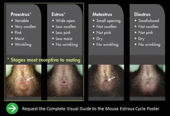
Download our “Visual Guide to the Mouse Estrous Cycle” poster for more reference images.
Cytological Identification of Estrous Cycle Stage. In contrast, vaginal smear analysis provides a more definitive method for determining estrous cycle stage. With this approach, estrous cycle stage is identified based on the relative abundance of 3 cell types: leukocytes, nucleated epithelial cells, and cornified epithelial cells. Proestrus is identified by the abundance of nucleated epithelial cells, while estrus is predominated by cornified epithelial cells (Quignon, Curr Protoc. 2024). However, this method is more time-consuming as it requires collection of vaginal cells and microscopic examination, with or without staining.
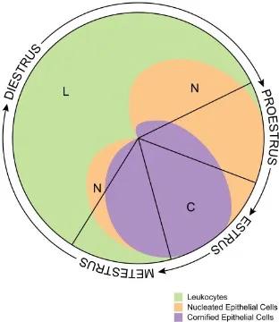
Byers et al., PloS One, 2012
Estrous Cycle Synchronization: Methods Reported in Literature
Theoretically, synchronization of the estrous cycle can contribute to the efficiency of establishing timed pregnancies while reducing the need for routine estrous stage assessment.
For those interested, several hormonal synchronization methods have been reported in the literature, represented by the progesterone-based approaches (Hasegawa et al., JRD, 2017; Sato et al., Reprod Med Biol. 2026) and luteinizing hormone-releasing hormone agonist -based approaches (Sari et al., Sci Rep. 2022). Estrous cycle synchronization has also been reported for application to generate pseudopregnant females for embryo transfer and in combination with superovulation protocols to increase oocyte yield (Hasegawa et al., BOR, 2016).
A traditional non-hormonal approach is the Whitten effect, in which exposure of group-housed females to male-associated pheromonal cues, commonly through male-soiled bedding, can help synchronize estrous cycling (Whitten, J. Endocrinol. 1956). However, this method is less precise than direct estrous stage assessment, and transfer of soiled bedding may pose biosecurity concerns. It should therefore be used only when appropriate and under institutional animal care guidance.
Checking for Vaginal Plugs
Females that mate during the dark phase typically have a copulatory plug at the vaginal opening the following morning. The plug is formed by coagulation of seminal vesicle secretions together with proteins derived from the coagulating gland and prostate, indicating successful copulation while also contributing to fecundity (Dean, PloS Genetics, 2013; Noda et al., BOR, 2018). Because vaginal plugs may become dislodged or difficult to detect over time, females should be examined early after the onset of the light phase. The optimal timing for plug checking depends on the light–dark cycle used in the animal facility and should be adjusted accordingly.
The presence and persistence of a vaginal plug can vary between strains. For example, plugs from C57BL/6J mice are often thin and may dissolve rapidly, whereas plugs in other strains may persist longer but can still be dislodged before dissolution. Despite these limitations, the presence of a copulatory plug remains the most widely used and practical indicator of successful mating for establishing timed pregnancies. In addition, a vaginal plug does not necessarily indicate successful pregnancy but only that copulation has occurred. Some males, particularly from strains such as BALB/cJ, may mate even when females are not in estrus. Therefore, confirming the estrous stage prior to pairing can improve the efficiency of timed mating and increase the likelihood of successful pregnancy.
Overall, timed mating supports the principles of the 3Rs in animal welfare by reducing the number of animals required for breeding and refining experimental precision by enabling the use of age-matched cohorts.